Current location:
Home
> News > Product News > Interactive Flat Panel
 Release date: 2026-09-28
 Clicks:0
From image display, partial magnification to annotation, case discussion and distance teaching, how to analyze Qtenboard help medical students from "seeing images" to "understanding images"
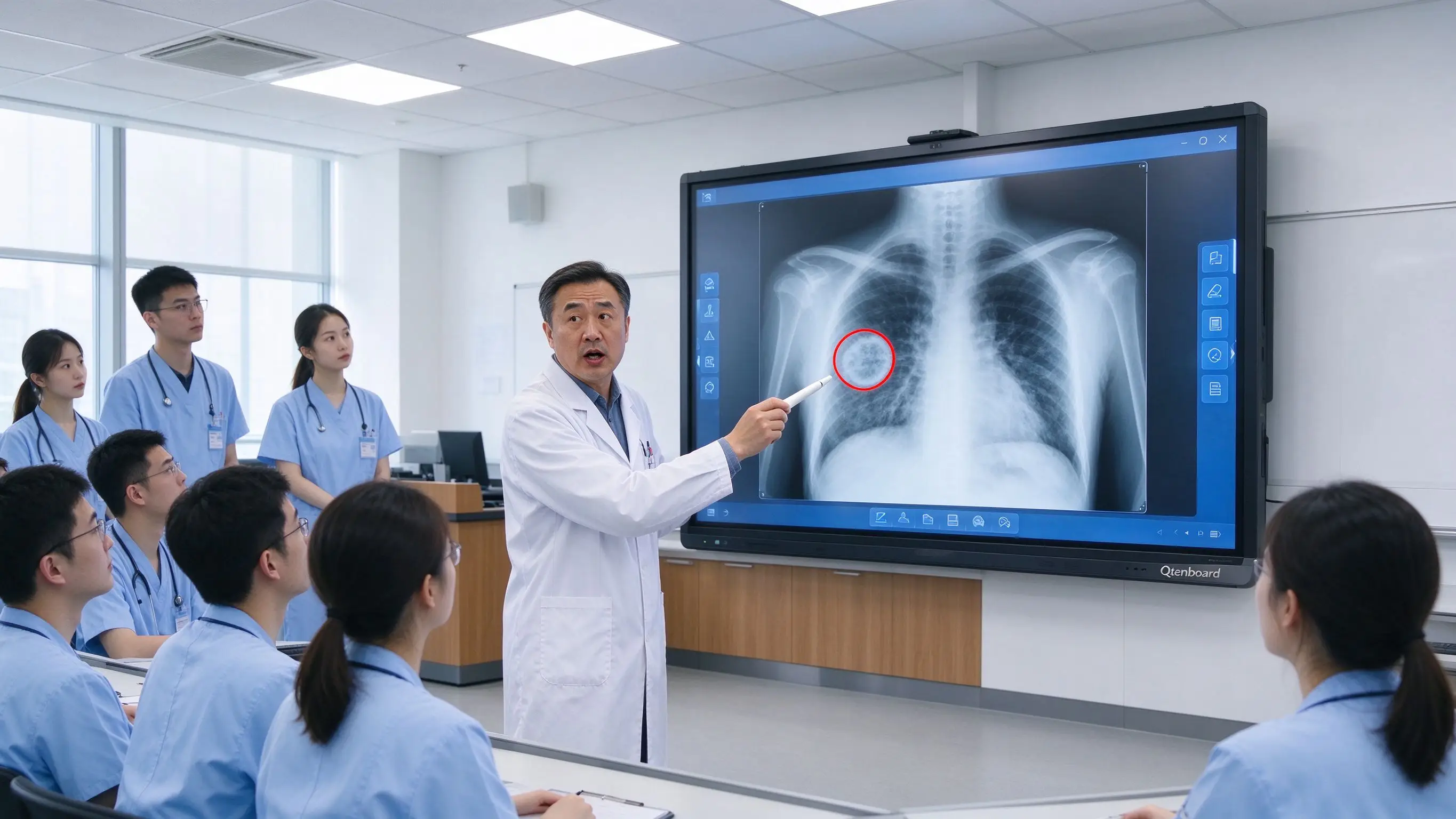
In traditional video teaching, the teacher opens the X-ray film on the computer, and the students watch from a distance, and some details are not clear. Teachers need to constantly zoom in, zoom out and switch windows. Images, anatomical maps, case data and operation videos are scattered in different files. Students are easy to remember "this is some kind of disease", but they do not understand the basis of judgment, and it is difficult to save the annotation and discussion results after the class ends.
Medical imaging teaching should not stop at "showing a film", but should help students establish a complete cognition from image features, anatomical location, clinical symptoms to judgment path. When the teacher can directly enlarge the suspicious area on the large screen, mark the fracture line or lung shadow on the image, and display the image side by side with the three-dimensional anatomical model, the image teaching can be upgraded from one-way display to joint reading, case reasoning and remote collaboration.
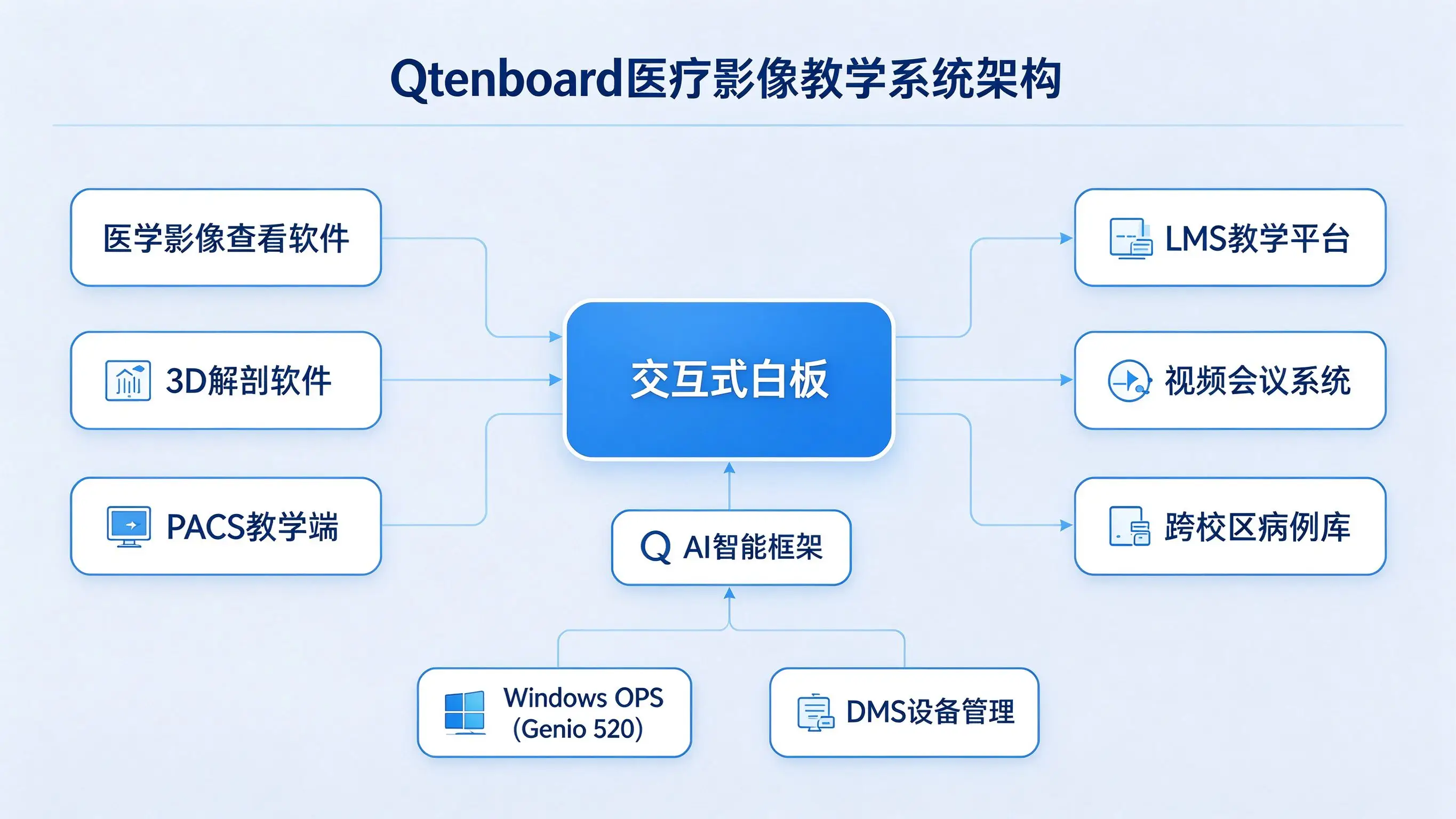
Qtenboard (Shenzhen Qunmao)Founded in 2005, we have been dedicated to interactive display manufacturing for 20 years. We operate a 330,000㎡ smart manufacturing industrial park in Shenzhen and Guangzhou, featuring eight fully automated production lines with a daily output capacity of 200 units. Our solutions serve 120 countries and regions worldwide, and we have provided services to over 15,000 corporate clients to date. The Qtenboard interactive whiteboard covers 55-110 inch full-size segments, is equipped with Q AI system-level framework and MediaTek Genio 520 platform, and has successfully served overseas medical colleges such as Kharkov National Medical University, providing a complete scheme from hardware to software for medical imaging teaching, radiology reading and clinical skills training.
The small-sized computer screen is not suitable for many people to watch. The projection is affected by ambient light. It is difficult for students in the back row to observe small structures. Complex cases need to be switched between multiple windows. In a typical video teaching scene, the teacher faces a 24-inch monitor, 20-30 students are crowded in the classroom, only 3-5 people in the front row can see the details of the fracture line or lung nodule, and the rest of the students can only make up for it through after-school materials.
It is difficult for students to correspond the two-dimensional slice to the three-dimensional structure, the change of image level is not easy to understand, and the relationship between blood vessels, bones, organs and lesions is difficult to present at the same time. Medical education research shows that 3D visualization technology is widely used to help medical students to establish spatial knowledge and structural relationship, but it should be combined with teachers' explanation, traditional atlas, specimens and clinical images for comprehensive teaching.
Teachers usually master the mouse operation, students can only watch passively, the discussion process is difficult to record, and the judgments of different students cannot be compared in parallel. The core of image teaching is to cultivate students' "reading thinking", which requires students to mark, explain and discuss on the image, rather than just watching the teacher's operation process.
Imaging instruction also typically involves symptoms, medical history, laboratory tests, imaging reports, surgery or treatment options, prognosis, and review results. If these contents are scattered in multiple systems, the teaching process is easily interrupted. Teachers often need to switch between PACS system, PPT, PDF report and paper materials repeatedly, which affects the continuity of teaching.
Remote students cannot see the details marked by teachers, remote teachers are difficult to guide students to locate, different colleges and universities use inconsistent versions of images and materials, and there is a lack of reusable duplicate files after the class.
Magnify fractures, shadows, nodules and organ boundaries, observe small blood vessels, bone structure and tissue changes, let the whole class see the area that the teacher is analyzing, and support partial screenshots, preservation and labeling. Qtenboard 4K UHD resolution and AG anti-glare glass ensure that the image details are clear and readable, and 20-50-point touch supports multiple people to mark at the same time. The touch accuracy is 1.5mm and the single-point response time is 7ms, ensuring the accuracy and real-time of the labeling operation.
X-ray film, CT slice, MRI image, ultrasound video, 3D anatomical model, case information, image report and clinical operation video can be displayed at the same time. The Qtenboard supports multi-window display and split-screen operation at the same time, and completes multi-source content comparison through the same screen.
Teachers or students can circle abnormal areas, mark anatomical structures, draw the scope of lesions, add arrows and text, record the basis for judgment, compare normal and abnormal images, and save annotated versions of different stages. The Q AI supports handwriting recognition and graphic recognition, and hand-drawn annotations can be quickly converted to standard graphics or digital content.
Locate from two-dimensional slices to three-dimensional regions, rotate the anatomical model, hide or show tissue layers, compare blood vessels, nerves, and bones, and interpret structural projections in the image. The Qtenboard Android Windows OPS dual system can run mainstream 3D anatomy software and medical image viewing tools, and the switching time is less than 3 seconds.
Students mark different judgments on the screen, the group numbers the abnormal areas, distinguishes different opinions by color, and the teacher focuses on comparing the results and discussing the causes of wrong judgments. Qtenboard support 20-50 touch, multiple students can operate on the large screen at the same time.
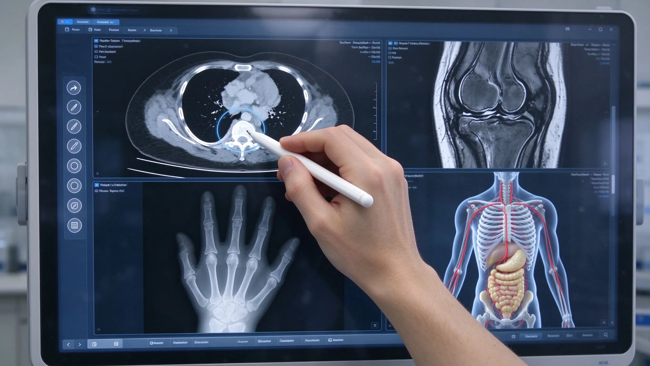
Teaching content:Bone structure, chest anatomy, joints and spine, normal imaging features, common abnormalities.
Qtenboard applications:Magnify the image, mark the anatomical landmarks, compare the normal and abnormal, let the students locate the lesion, and make structural matching and case judgment. In the application of Kharkov State Medical University, the large 75-inch 4K screen ensures that students sitting in the back of the classroom can also clearly see the details of the image.
Teaching content:Multi-plane image, different levels of switching, tissue and organ contrast, lesion location, image sequence differences, three-dimensional reconstruction.
Qtenboard applications:Show serial sections, compare different levels, mark the scope of the lesion, connect the image with the three-dimensional model, and discuss the diagnostic ideas. The Windows OPS module runs professional image viewing software and supports DICOM data import and display.
Teaching content:Real-time video, probe orientation, anatomical changes, blood flow and dynamic observation, operator manipulation.
Qtenboard applications:Play the ultrasound video, pause and label, zoom in on the local area, explain the probe angle, show the standard image and common errors. Support external ultrasonic equipment signal input.
Teaching content:Trauma imaging, fracture, pneumothorax, hemorrhage, foreign body, acute abdominal imaging.
Qtenboard applications:Quickly identify hazard signs, prioritize, build time awareness, discuss next inspection or treatment process, combine team communication and first aid simulation. Q AI meeting intelligence automatically records teaching timelines and key decision nodes.
Lesion location, tissue boundary, organ relationship, surgical approach, risk area, treatment plan. Teaching presentations are not a substitute for formal clinical planning systems, real patient data must be de-identified, and all clinical judgments should be made by qualified professionals.
The left side shows the CT slice, and the right side shows the three-dimensional organ model. The teacher establishes the corresponding relationship by marking, and the students judge the spatial position according to the slice. The Qtenboard supports multi-window display, where images, models, and case information can be presented simultaneously on the same screen.
Skin, muscle, bone, blood vessel, nerve, viscera, lesion. Through the Hide, Show, and Transparent modes, students can see what structures in the body the "shapes in the image" correspond.
Show the transverse, coronal, and sagittal planes, mark the slice direction, explain the appearance of the lesion in different planes, and discuss the puncture, surgical, or catheter path.
The patient's symptoms are given, relevant images are shown, students mark possible structures involved, teachers explain the relationship between anatomy and symptoms, and save the conclusions of the case discussion.
Compare the image with the report text, let the students find out the corresponding structure of the report, discuss whether the description is accurate, learn the professional terminology of medical imaging, and train the ability of structured expression.
Hide the structure name, students touch positioning, say the structure name and function, teachers add clinical significance. Qtenboard supports 20–50-point touch, allowing multiple students to interact on the large screen simultaneously.
Provide normal and abnormal images, students circle the differences, the group explains the basis of judgment, and the teacher shows the standard analysis path. Q AI supports graphic recognition, and hand-drawn annotations can be converted to standard graphics.
History, symptoms, and images are presented, students develop diagnostic hypotheses, label evidence on images, compare different interpretations, and discuss what information is needed next.
Students complete structured descriptions, teachers modify the statements on the whiteboard, discuss terms, locations, and degrees, and export the results of the course exercises. Qtenboard supports saving whiteboard pages and annotations with one click.
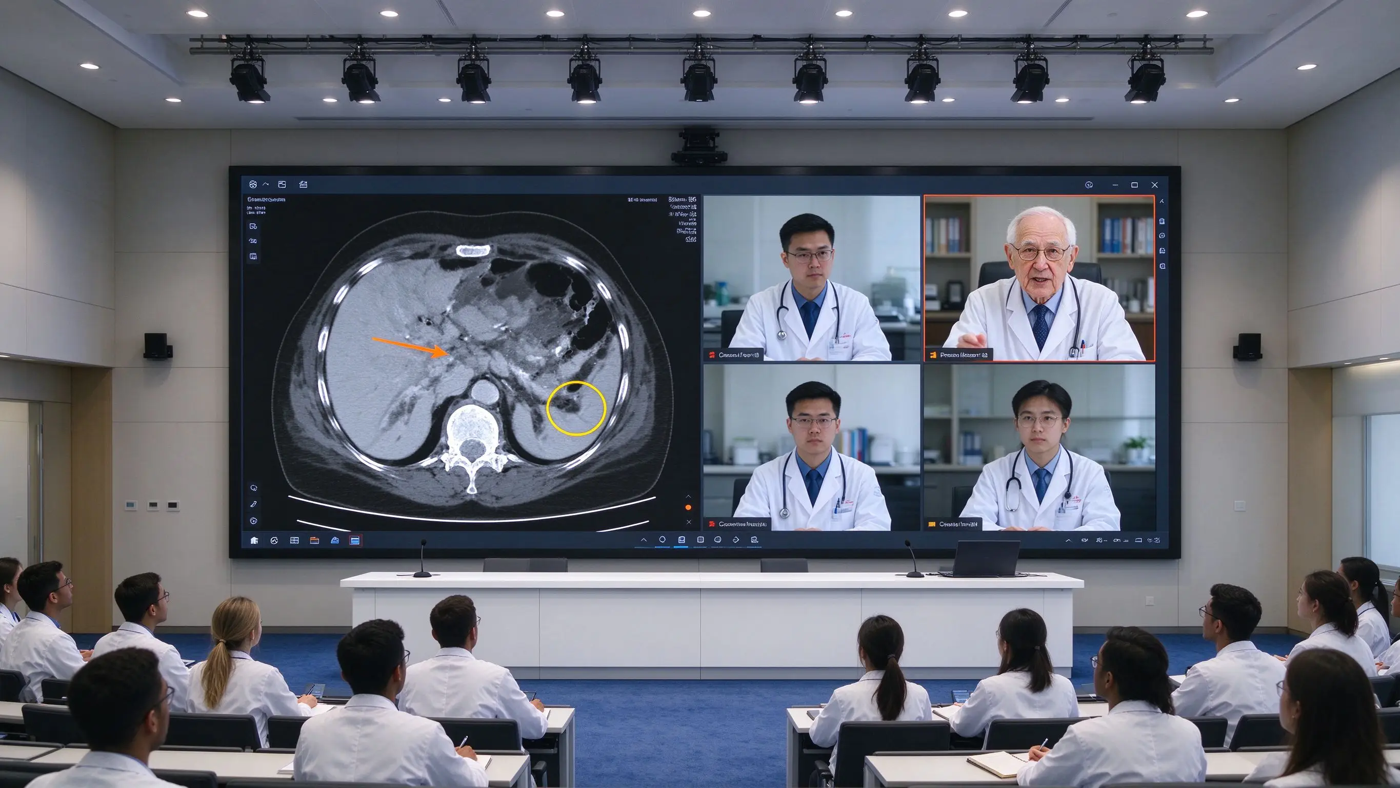
Remote teachers share images, students mark on the whiteboard, different colleges and universities compare and judge, experts comment and save annotations, forming a cross-campus case library. The Qtenboard supports up to 9 devices at the same time, and the Q AI supports real-time translation and real-time subtitles in 166 languages.
Qtenboard DMS supports remote update, account and permission management, application deployment, network monitoring, software version control, operation log, and data backup. For medical schools with multiple imaging classrooms and training centers, DMS can centrally manage all equipment and reduce IT operation and maintenance workload.
Name, hospital number, date of birth, contact information, facial or other identifiable information, examination time, and relevant metadata should be processed before teaching the image. Q-AI core transcription runs locally on the device and can be used without networking, reducing data privacy risks at the architectural level.
Teaching whiteboard is not a clinical diagnostic workstation, should not be unauthorized to modify the original data of PACS, should not be written into the official patient file without audit, teaching equipment should not be used as a clinical medical order system. If the project involves clinical diagnosis, it must be specially confirmed by the hospital imaging department, IT department and compliance team.
Q AI is not a cloud-based plugin; it is a native intelligent architecture deeply integrated into the hardware, software, and algorithm layers. Core transcription runs locally on the device and can be used without an internet connection. In image teaching, Q AI supports handwriting recognition and graphic recognition, and the annotation drawn by teachers can be quickly converted to standard graphics. Q AI also supports real-time translation in 166 languages. For medical schools that accept international students, the imaging terms explained by teachers in Chinese can be translated into English in real time.
The Qtenboard Genio 520 interactive tablet platform is based on the MediaTek Genio 520 (MT8371) chip, and the 8th generation MediaTek NPU is directly provided on the interactive tabletDedicated AI acceleration for 10 TOPS. In video teaching scenarios, this means real-time handwriting recognition, shape correction, and gesture control all run on the device, with zero internet dependency, zero cloud latency, and zero data privacy risk.
Based on TSMC's 6nm FinFET process, the Genio 520 achieves high performance with low thermal output, which enables QtenboardFanless interactive tablet designMake it possible-essential for demanding acoustic environments such as imaging classrooms and medical simulation centers. A fanless design means zero noise, no moving parts, and no maintenance schedule. MediaTek’s official commitment to the Genio 520 is10 year standard supply life cycle(EOL 2035), eliminating the risk of obsolescence that consumer-grade SoCs usually phase out in 2-3 years.
| Size | Typical teaching scene | Qtenboard configuration |
|---|---|---|
| 65/75 inches | Small teaching group, video display classroom, basic reading film | 4K, 20-50 touch, Android 14 |
| 86 inches | Standard imaging classroom, radiology teaching | 4K/5K, dual system, 20-50 touch |
| 98/105/110 inches | large class teaching, medical simulation center, remote imaging teaching | 4K, high brightness, independent OEM production line |
Dimensions should be identified in conjunction with classroom depth, number of students, image detail, and viewing distance. Qtenboard services are available in 120 countries and regions, with after-sales response times of ≤4 hours.
Kharkiv National Medical University is located in Ukraine and is a well-known medical education institution in Eastern Europe. In the process of education modernization, the school made a forward-looking procurement decision-to introduce 10 sets of Qtenboard 75-inch interactive flat-panel displays, equipped with sliding green boards, for the teaching of core courses such as imaging and anatomy.
The 4K ultra-high definition resolution Qtenboard the large 75-inch screen ensures that the fine structure of X-rays, CT slices and MRI images is presented in extremely fine image quality, and students can capture every key detail. High-brightness and high-contrast screen technology ensures that images are readable in different lighting conditions, making it easy for students to see the content even in the back of the classroom.
The touch functionality greatly enhances classroom interactivity. In the imaging course, teachers can use touch operation to accurately point out the fracture line or lung shadow on the X-ray film, and students can also operate on stage and mark their own judgment on the image. Teachers’ handwriting and annotations can be saved in real time, making it easy to organize digital lesson plans after class and share them with students for review.
The image teaching center of a medical university used the traditional configuration of projector and computer before. When the teacher demonstrated the image software, the students could only watch it. The students in the back row could not see the details clearly and the students could not directly operate the image.
After deploying Qtenboard 86-inch interactive whiteboard, teachers can zoom in on X-ray, CT and MRI slices on a large screen to directly mark suspicious areas. The students take turns to operate on the stage, circling the abnormal structure on the image, and the teacher focuses on comparing the judgments of different students. The Qtenboard-supported structured editing function of blackboard writing allows teachers to quickly adjust the word order of text through hand-drawn displacers, making it easy to organize imaging terms and notes. Windows OPS module runs professional image viewing software smoothly, and the switching time is less than 3 seconds. Classroom engagement has significantly improved, and teachers have reported a noticeable increase in the frequency of questions asked by students seated in the back rows.
A provincial hospital needs to provide imaging teaching training to five grass-roots hospitals. There is a lack of radiology experts in grass-roots hospitals, and experts cannot teach on-site for a long time.
After the deployment of Qtenboard 75-inch interactive whiteboard, the central hospital will conduct remote demonstration through video conference and whiteboard. The branch students will simultaneously view the images, anatomical models and case data, annotate and guide the remote experts, and save the course and resume the contents after class. Q AI supports real-time subtitles in 166 + languages, the standardization of cross-hospital training has been significantly improved, and the travel cost of experts has been reduced by about 70%.
Requirements for instructional displays and clinical diagnostic displays may differ. Correct approach: clear equipment use, and follow the hospital imaging department and regulatory requirements. The Qtenboard is positioned as a teaching and collaboration tool that complements rather than replaces clinical diagnostic displays.
Without proper software, whiteboard cannot undertake multi-plane, three-dimensional and case teaching. The software platform and license should be confirmed first. Qtenboard Windows OPS supports mainstream medical image viewing software.
Hospital data policies must be de-identified and adhered to, and data processing, storage, and permissions need to be clarified. Q AI core transcription runs locally, reducing data privacy risks.
Students are unable to build true image judgment. Structure location, anomaly labeling, case reasoning and group discussion should be designed. Qtenboard support 20-50 point touch, multiple students can operate at the same time.
Too strong anti-glare may affect image detail. Should be tested by real X-ray, CT and MRI samples. Qtenboard AG anti-glare glass reduces reflections while maintaining sharp image detail.
Projects also include OPS, software, camera, audio, installation, training and data security. A full TCO assessment should be used.
Can interactive whiteboards be used directly for clinical diagnosis?
Plain interactive whiteboards are primarily intended for teaching, case discussion, training, and remote collaboration and should not be a default replacement for certified clinical diagnostic monitors or PACS workstations. If the project involves clinical diagnosis, it must undergo a dedicated review and approval process involving the hospital’s radiology department, IT department, and compliance team. The Qtenboard is positioned as a teaching and collaboration tool, complementary to the clinical diagnostic display.
What is the size of medical imaging teaching?
Small teaching groups may consider 65 or 75 inches; large classes, distance learning and medical simulation centers may consider 86 or 98 inches. Final confirmation should be combined with image details, seating distance, number of people and spatial layout. Qtenboard covers the entire size range from 55 to 110 inches and can provide recommendations for selecting the appropriate size. Kharkiv State Medical University selected 75 inches for the standard imaging teaching classroom.
Do I need to Windows OPS?
If only basic images, PPT and videos are displayed, the Android may meet the requirements. If you need to run professional imaging software, 3D models or Windows teaching platforms, you should evaluate Windows OPS or external high-performance computers. Qtenboard OPS module based on Genio 520 platform, optional i5/i7 configuration, support mainstream medical image viewing software.
Can I connect to PACS?
Technical validation is required based on PACS version, network structure, authority, and hospital security policy. It is recommended to use de-identified teaching data for testing first, and not to directly access the clinical core environment without approval. Qtenboard Windows OPS supports professional medical imaging viewing software, and its local operation does not affect the hospital’s network security architecture.
Can Qtenboard customize the medical teaching system?
Logo, language, startup interface, software entry, OPS, camera, microphone, stand and device management configuration can be evaluated according to the project. A dedicated OEM production line ensures that customization does not affect the delivery schedule of standard products; the lead time for custom orders is extended by only 3–5 days. The CKD/SKD local assembly model helps partners reduce tariff costs and meet localization requirements in government procurement.
Does the Qtenboard equipment support the unified management of multiple hospitals?
Support. Qtenboard DMS (Device Management System) supports global multi-classroom unified monitoring, batch update and permission management. IT teams can manage all video teaching devices in one background, supporting remote power control, OTA firmware updates, batch application distribution, and AI-driven predictive diagnostics. The AI-driven predictive diagnostic module checks hardware parameters such as panel temperature, internal voltage and color values, and provides early warning to avoid unexpected downtime of the equipment during teaching.
If you are planning a medical imaging classroom, a radiology teaching center, or a remote imaging teaching program
The Qtenboard provides 55-110 inch full-size interactive whiteboard, Q AI system-level framework (166 language, local operation, 5-second minutes), Genio 520 platform (10 TOPS device-side AI, fanless mute design), DMS device management and OEM/ODM/CKD/SKD customization services.
Get a guide to the selection of Qtenboard medical imaging teaching solutions, or book a factory visit and prototype test.
 Partner Links:
 en
 中文

 Previous:
 Return to list